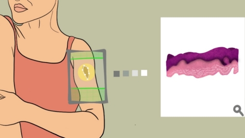
The next time you have a suspicious-looking mole on your back, your dermatologist may be able to skip the scalpel and instead scan the spot with a noninvasive “virtual biopsy” to determine whether it contains any cancerous cells. Similarly, surgeons trying to determine whether they have removed all of a breast tumor may eventually rely on an image captured during surgery rather than wait for a pathologist to process the excised tissue.
Stanford Medicine researchers have developed a method that uses lasers to penetrate tissue and create a high-resolution, three-dimensional reconstruction of the cells it contains. From this virtual reconstruction, they can make cross-sectional images that mimic those generated by a standard biopsy, in which a sample of tissue is sliced into thin layers and placed on a slide to be examined under a microscope.
[Read more at Stanford Medicine News Center]


